History of Bionic Vision
Advances in medicine, surgery and electronics have set the stage for a fusion of the physical and biological sciences; one in which visual prostheses may offer a form of artificial vision to people who are blind. For excellent reviews on the subject, see [1, 2, 3].
The field has not progressed in a straight line. It has advanced in waves: bursts of intense research, followed by quieter periods and renewed interest. Cortical, retinal, and other approaches have developed side by side rather than replacing one another. The sections below are editorial. They mark shifts in emphasis, not dates on which any line of research began or ended.
Research Waves at a Glance
Visual prosthesis research has advanced in bursts rather than a straight line. Cortical work peaked in the 1970s, slowed, and has returned strongly since 2016; retinal implants rose in the 2000s, stumbled commercially around 2019, and are being renewed by a new generation of devices.
Discovering Electrical Vision
1750s to 1950s
Long before anyone tried to build a visual prosthesis, scientists learned that electricity could produce visual sensations. The end of the 18th century saw the introduction of electrophysiology as a scientific discipline, opening the door to many studies of brain stimulation. In the early 20th century, neurosurgeons treating epilepsy and battlefield head wounds began to map the phosphenesthe perception of light without light entering the eye evoked by stimulating the visual cortex.
1752
Benjamin Franklin (opens in a new tab) theorized to the Royal Society of London that sight and hearing could be restored with the use of electricity [4].
1755
Charles Le Roy (opens in a new tab), an 18th century French scientist, made the first known attempt to restore sight through electrical stimulation (presumably after reading Benjamin Franklin's theory). Le Roy was able to evoke flashes of light (phosphenesthe perception of light without light entering the eye) and other visual disturbances in a blind patient by sending electrical currents through a wire wrapped around the patient's head [5].
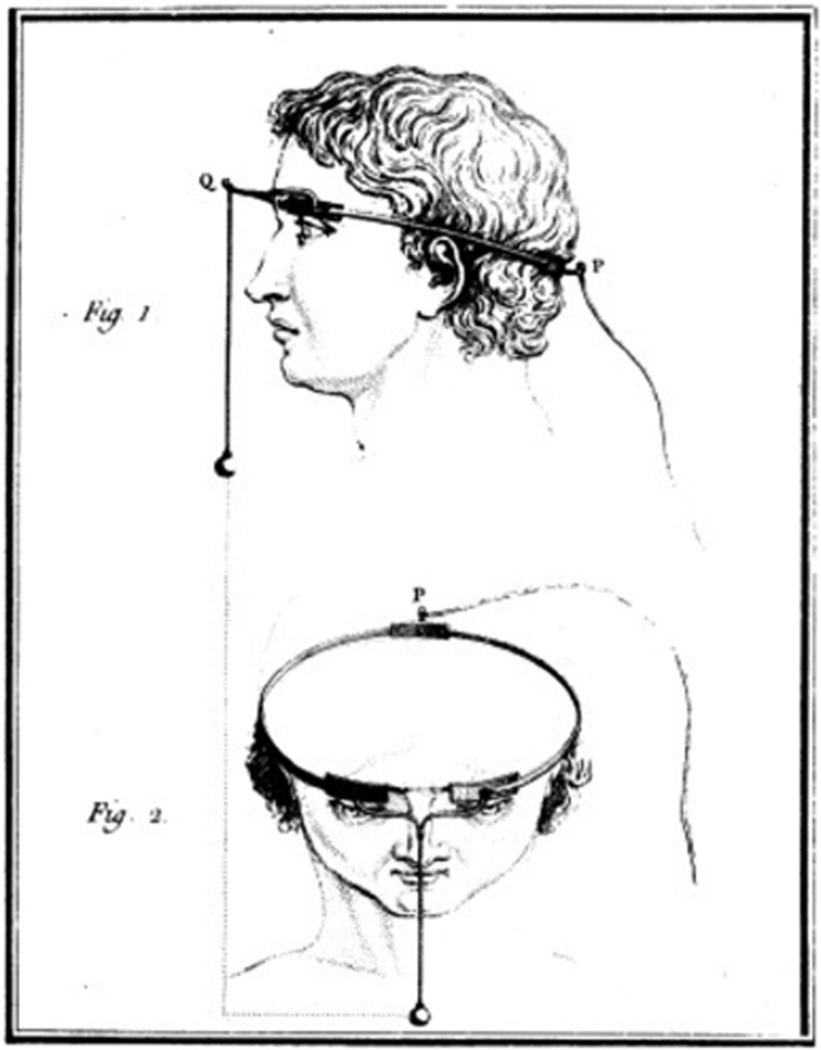
Fig. 1: Method used by Charles Le Roy to evoke visual disturbances in a blind volunteer. A wire was wrapped around the patient's head with current applied through it. (reproduced with permission from [6]) 1870
Two German physicists called Gustav Theodor Fritsch (opens in a new tab) and Eduard Hitzig (opens in a new tab) performed a number of experiments involving systemic stimulation across the cerebral hemispheres of dogs. These studies demonstrated that cerebral hemispheres were excitable at a time when many scientists believed that they were not [7].
1884
Victor Horsley (opens in a new tab), a British scientist, is credited as the first to use electrical stimulation of the cerebral cortexthe folded outer layer of the brain involved in perception, thought, and voluntary action intraoperatively. Horsley electrically stimulated an occipital encephalocele in a 6-week old child [8], in response to which the child's eyes showed rapid conjugate deviations.
1918
Kurt Löwenstein & Moritz Borchardt electrically stimulated the exposed visual cortex of a patient who had persistent seizures from a bullet wound on the left side of his head. The stimulation produced flickers of light that closely resembled the visual phenomena the patient would see preceding a seizure, showing that stimulation of the visual cortex could produce point sources of light [9].
1924
Fedor Krause (opens in a new tab) introduced surgical operations for treatment of epilepsy in Germany, performing over 400 operations on patients with epilepsy. When he stimulated the cortex of a patient with a gunshot wound, the patient reported seeing jagged rings of light and stars in different patterns. Krause observed that even though his patient was hemianopicloss of vision in one half of the visual field for nine years, he still experienced visual hallucinations in his blind hemifield preceding a seizure. Electrical stimulation could provoke similar visual imagery in the blind hemifield as well [10].
1929
Otfried Förster (opens in a new tab), a German neurologist and neurosurgeon, examined a patient with a projectile injury that resulted in occipital seizures. He demonstrated that stimulation of the occipital polethe rear part of the cerebral cortex that contains most of the visual cortex and its surrounding cortex produced phosphenes that were motionless and located at the center of the patient’s visual field [2].
1947
The American-Canadian neurosurgeon Wilder Graves Penfield (opens in a new tab) examined 17 years of visual cortex stimulation in about 330 operations. He stimulated a wide area of the visual cortex and also one area outside the occipital lobe, which still produced visual imagery. The observations of Penfield were unique in that patients reported seeing phosphenes of different colors [11].
The First Wave: Cortical Visual Prostheses
1958 to 1982
By the 1950s, it was well known that electrical stimulation of the occipital poles could produce punctate phosphenes in the center of the visual field [3]. This knowledge, plus Krause's observation that it was possible for a blind patient to perceive visual imagery upon stimulation of the occipital cortex, prompted the suggestion that a visual prosthesis might be a practical possibility.
What followed was the first sustained effort to build one, with the goal of creating patterned artificial vision that would let recipients read printed text. Government funding carried the wave through the 1970s. The year Brindley & Lewin published their groundbreaking paper, the UK Medical Research Council formed the Neurological Prostheses Unit, with Brindley as its director. In the US, the University of Utah started a sensory prosthesis research program with William Dobelle as director, and the National Institutes of Health (NIH) established a neuroprosthetic program at the US National Institute of Neurological Diseases and Blindness (NINDB, now known as NINDS). By the early 1980s, momentum had faded. The implants placed large electrodes on the surface of the brain, needed strong currents, produced coarse phosphenes, and carried risks of infection and seizures.
1958
"I see a flash!" - Button (1958, p.54)
John C. Button and Tracy Putnam at Cedars of Lebanon Hospital (now Cedars-Sinai) in Los Angeles created and implanted the first ever bionic vision device in a 36-year old woman who had been completely blind for 18 years. Two pairs of stainless steel wires were implanted into the cortex and connected to a simple stimulator. The patient reported seeing vivid flashes of light and (with the use of a photocell) was able to detect the presence and relative brightness of a light source [3].
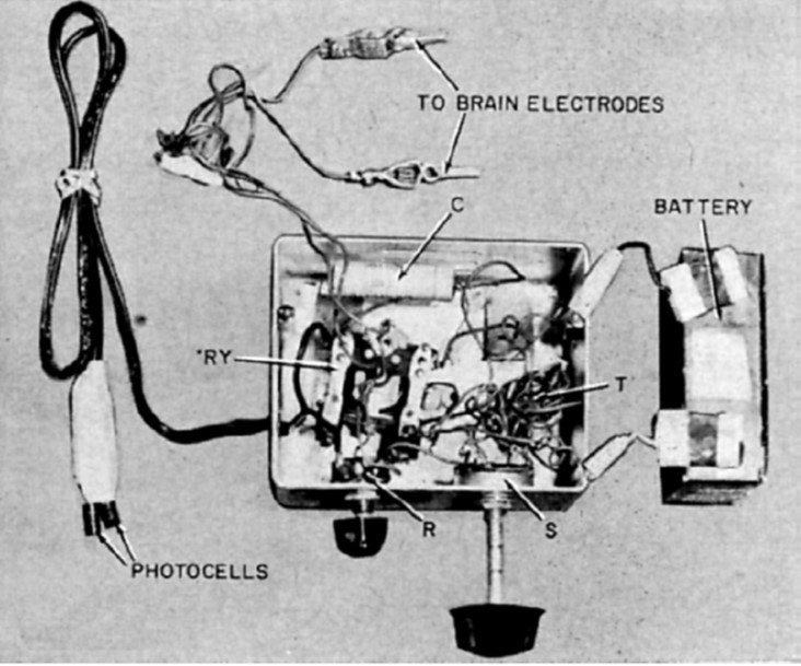
Fig. 2: Button's stimulating apparatus was connected to an array of four stainless steel wires implanted in the occipital cortex of a blind woman. (reproduced under CC BY-NC-ND 4.0 (opens in a new tab) from [2]) 1965
Giles Brindley, a physiologist at the University of Cambridge, had been undertaking a program of research to develop a fully implanted, multielectrode, wireless visual cortex stimulation device, with the goal of allowing prosthesis recipients to read printed or handwritten text [2].
1966
The first Conference on Visual Prosthesis was held in Endicott House at the Massachusetts Institute of Technology. The conference chair Theodore Sterling explained that after initial difficulty in bringing together potential participants, the conference was finally held under the proviso that the proceedings would not be made publicly accessible [2].
1968
"like a star in the sky" (in the center), "...more like a cloud" (in the periphery) - Brindley & Lewin (1968)
Brindley and Lewin implanted their first cortical implant that consisted of an array of 80 small, square, platinum electrodes embedded in a silicone cap. Each electrode was hard-wired to a receiving coil that was part of an array of coils also embedded in silicone and implanted under the scalp. 39 electrodes in the implant produced phosphenes across the patient’s visual field when stimulated. Centrally evoked phosphenes were small and flickering whereas peripherally evoked phosphenes differed more in size, shape, brightness, and sharpness [12].
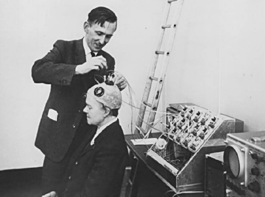
Fig. 3: Brindley stimulating the visual cortex of his first implant recipient, placing a stimulating coil over a matched receiving coil implanted under her scalp. (reproduced under CC BY-NC-ND 4.0 (opens in a new tab) from [2]) 1969
The second Conference on Visual Prosthesis was held at the University of Chicago, aimed at trying to find future directions necessary to explore a possible visual prosthesis [2].
1974
"a colour from another world" - Dobelle & Mladejovsky (1974)
William Dobelle and his group developed a removable multielectrode cortical surface array, which was tested on 36 sighted patients undergoing occipital surgery [13] and 2 blind volunteers who had been blind for 7 and 28 years, respectively [14]. The array contained 64 hexagonally arranged platinum discs placed only over the mesial occipital cortex, each of 1-mm2 surface area. Their results in blind subject closely agreed with Brindley & Lewin's [12], noting that phosphenes could be elicited with stimulation of V2 and persisted with suprathreshold stimulation. The same was not true in sighted volunteers, where phosphenes did not always flicker and were occasionally colored, always disappeared immediately upon cessation of stimulus, and gradually faded with continuous pulse trains [13].
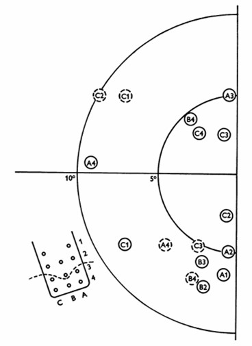
Fig. 4: Phosphene map in the visual field for case no. 36. Phosphenes indicated by dashed circles appear only at high amplitudes. The electrode array and numbering system are also shown, along with a dashed line showing the postulated position of the calcarine fissure. (reproduced with permission from [14]). 1974
Over the same period, researchers from the Huntington Institute of Applied Medical Research also developed and tested a cortical array comprising smaller, "strip" arrays of 18 platinum surface electrodes, placed separately on both the occipital pole and mesial occipital cortex of sighted patients [2, 15]. The implant was able to elicit phosphenes, but could also cause seizures, which led the NIH to abandon the group's effort in favor of research on "safe and effective methods for neural stimulation" [2].
1975
Another group funded by the NIH and based at the Massachusetts General Hospital in Boston had been undertaking intraoperative visual cortex stimulations on five patients throughout the early 1970s. However, only three patients reported seeing phosphenes, with the two failures attributed to postoperative swelling and patient drowsiness [2].
1978
Dobelle and colleagues continued to improve their implant design throughout the 1970s, adding a transcutaneous connectora connector that passes through the skin to link an implanted device with external equipment to permit chronic implantation of electrodes and spectacle-mounted cameras. One of Dobelle's early patients retained his implant for over 20 years, with no reported complications of infection or seizure [2].
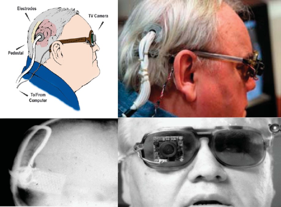
Fig. 5: Different views of a recipient of the Dobelle implant, first implanted in 1978. In 2000, his external hardware & software was upgraded, including the miniature camera shown, greatly enhancing the functionality of his prosthesis. (reproduced with permission from [6]) 1982
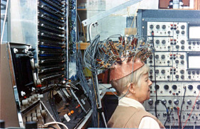
Fig. 6: "Miss Bonnett", recipient of the last visual cortex implant built by the MRC Neurological Prostheses Unit. (reproduced under CC BY-NC-ND 4.0 (opens in a new tab) from [2])
New Targets, New Electrodes
1983 to 2001
The 1980s and 1990s were a quieter period for cortical implants, but not a dormant one. Two lessons from the first wave shaped what came next. First, surface electrodes needed large currents and produced coarse phosphenes. This motivated penetrating microelectrodes that stimulate small groups of neurons from within the cortex. Second, the risks of brain surgery led researchers to look for other places along the visual pathway to stimulate: the retinathe light-sensitive neural tissue at the back of the eye that begins visual processing and the optic nervethe bundle of retinal ganglion cell axons that carries visual signals from the eye to the brain. These programs did not replace cortical research. They ran alongside it, and in 1996, landmark results for both intracortical and retinal stimulation were published within the same year.
1990
"two blobs fusing", "like a teddy bear" - Bak et al. (1990)
Martin Bak, Edward Schmidt and colleagues at the NIH reported visual sensations produced by intracorticalan implant with electrodes that penetrate into the cerebral cortex microstimulation of the human occipital cortex, using fine microelectrodes inserted into the cortex of patients undergoing surgery rather than discs resting on its surface [18].
1996
Building on that work, Schmidt and colleagues implanted 38 penetrating microelectrodes in the visual cortex of a 42-year-old woman who had been totally blind for 22 years. Over four months, 34 of the electrodes elicited phosphenes, at currents as low as 1.9 µA, far below what surface electrodes required. Electrodes spaced 500 µm apart produced separate phosphenes, a two-point resolution about five times finer than typically achieved with surface stimulation [19]. The study remains a landmark demonstration of what intracortical stimulation could offer a visual prosthesis.
1996
In the same year, Mark Humayun, Eugene de Juan and colleagues at Johns Hopkins showed that local electrical stimulation of the retinal surface in patients with outer retinal diseases resulted in focal light perception in patients who had been blind for many years [20]. Because the retina's output neurons survive in diseases such as retinitis pigmentosaa group of inherited retinal diseases that progressively damage photoreceptors, the eye offered a target that avoided brain surgery altogether. Joseph Rizzo and John Wyatt's Boston Retinal Implant Project ran similar short-term tests during eye surgery [21].
1998
Claude Veraart and colleagues in Brussels chronically implanted a self-sizing spiral cuff electrode around the optic nerve of a blind volunteer with retinitis pigmentosa. Stimulating the nerve produced localized visual sensations distributed throughout the visual field, establishing the optic nervea visual prosthesis that stimulates the optic nerve to create visual percepts as a third possible target [22].
2000
Alan and Vincent Chow's company Optobionics began implanting the Artificial Silicon Retina, a 2-mm subretinal chip with about 5,000 light-powered microphotodiodes, in people with retinitis pigmentosa. It was one of the first retinal implants designed to remain in the eye. Participants reported visual improvements, but some appeared far from the implant, suggesting a general protective effect on the retina rather than artificial vision [23].
2000
Dobelle reported connecting a digital video camera and computer to the cortical implant of a blind volunteer, the culmination of a development effort his group had begun in 1968 [24]. The report was a coda to the first wave of cortical prostheses: the system still relied on surface electrodes implanted more than two decades earlier.
The Rise of Retinal Prostheses
2002 to 2015
In the 2000s, the center of gravity moved to the retina. Retinal implants could be placed using established eye surgery and were aimed at people who had lost their sight to outer retinal degenerations. Human trials moved from brief intraoperative tests to chronic implants and, by 2013, to the first commercial approvals in Europe and the United States. Cortical research did not stop. Groups led by Richard Normann in Utah, whose Utah electrode array would later be used in several human trials, Philip Troyk in Chicago, Eduardo Fernández in Elche, and Arthur Lowery in Melbourne kept refining penetrating electrode arrays and wireless stimulators in the laboratory, laying the groundwork for the next wave.
2002
The first Argus I implants, developed by Mark Humayun's team at the University of Southern California with Second Sight, marked the start of chronic clinical testing of epiretinallocated on the inner surface of the retina, next to the vitreous prostheses. The first recipient, who was completely blind, saw spots of light on all 16 electrodes of the array, and with a camera could detect motion and recognize simple shapes [25].
2010
Eberhart Zrenner and colleagues in Tübingen reported results from an active subretinallocated beneath the neural retina, near the photoreceptors and retinal pigment epithelium microchip containing 1,500 light-sensitive photodiodes. Three previously blind participants could locate bright objects. One could name objects such as a fork or a knife, and read large letters and combine them into words [26].
2011
Argus II, an epiretinal implant developed by Second Sight Medical Products, received approval for commercial use in the European Union (CE Marka marking indicating that a medical device meets applicable European requirements). The implant was initially available at a limited number of clinics in Switzerland, France, and the United Kingdom, at an EU market price of $115,000 [27].
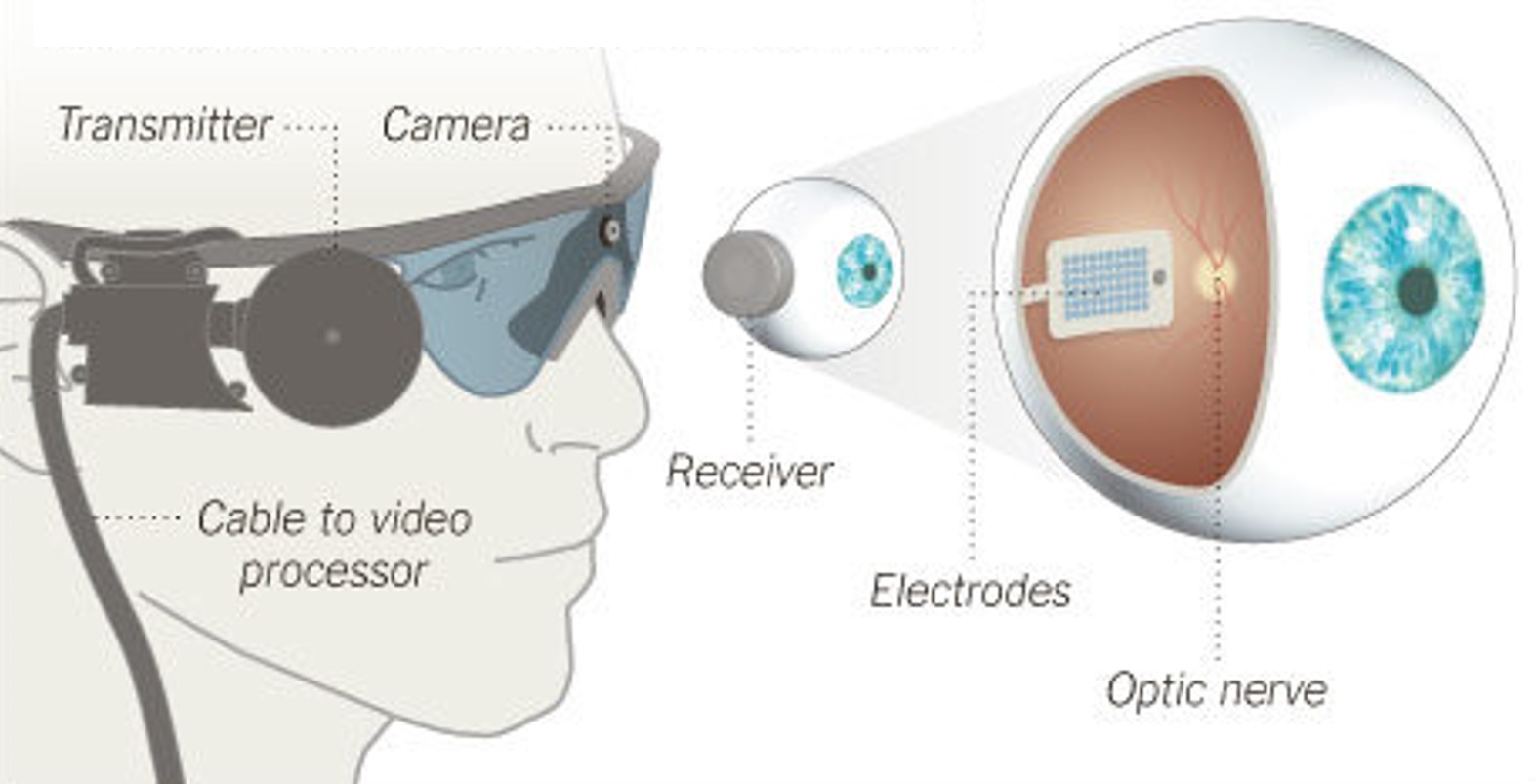
Fig. 7: Argus II Retinal Prosthesis System (Source: Second Sight). Camera images are processed and transferred to electrodes implanted in the back of the eye. 2012
Bionic Vision Australia, a national research consortium, implanted a 24-channel prototype in three people with end-stage retinitis pigmentosa. Unlike epiretinal and subretinal devices, its electrode array sat in the suprachoroidallocated between the choroid and sclera at the back of the eye space, between the sclera and the choroid, a location chosen to make surgery simpler and safer and to preserve any remaining vision. The trial, led by surgeon Penelope Allen at the Centre for Eye Research Australia, reported that the surgery was successful and the approach safe over twelve months of follow-up [28]. In Japan, Takashi Fujikado's group at Osaka University had reported a related approach, suprachoroidal-transretinal stimulation, in two patients in 2011 [29].
2013
Alpha IMS, the subretinal implant developed by Retina Implant AG, received the CE Mark as well [30]. The implant was initially available at around $130,000.
Meanwhile in the US, Argus II became the first retinal implant to receive FDA approval [31].
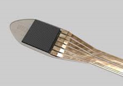
Fig. 8: Alpha IMS, a subretinal implant consisting of a 3x3mm2 microchip with 1,500 electrodes (Source: Retina Implant AG).
The Modern Wave: Returning to the Cortex
2016 to today
Since 2016, the cortex has returned to the center of the field, this time alongside retinal research rather than instead of it. Penetrating microelectrode arrays proven in other brain–computer interfaces, wireless implanted electronics, and a better understanding of how the visual cortex maps the visual field made new human trials possible. At the same time, first-generation retinal implants met a commercial reckoning, while a new generation of retinal devices and biological approaches such as optogeneticsthe use of light-sensitive proteins to control genetically targeted cells emerged. Recent work has shifted from evoking isolated phosphenes toward shaping coherent percepts, increasingly guided by recordings of the brain's own response to stimulation.
2016
2018
The first participant received the complete Orion system, including camera glasses, at UCLA. This marked the start of an early feasibility studya small, early clinical study of a medical device focused on initial safety and functionality that would enroll six participants at UCLA and Baylor College of Medicine [33].
2019
First-generation retinal implants went into commercial retreat. Second Sight discontinued Argus II to focus on Orion, and Retina Implant AG ceased business, citing Europe's regulatory climate and unsatisfactory results in patients. In 2020, Second Sight laid off most of its staff and later merged with Nano Precision Medical to form Vivani Medical, leaving the more than 350 Argus II recipients worldwide with little support for their devices [34].
These were serious setbacks despite earlier clinical and regulatory successes. The visual benefit the devices provided was modest, and the business case for sustaining them proved fragile.
2020
Two studies moved beyond isolated phosphenes. Michael Beauchamp, Daniel Yoshor and colleagues traced letter shapes on the surface of the visual cortex by stimulating electrodes in a dynamic sequence. Sighted and blind participants recognized the shapes accurately, blind participants at up to 86 forms per minute [35]. Pieter Roelfsema's group at the Netherlands Institute for Neuroscience implanted a 1,024-channel interface in the visual cortex of sighted monkeys. By stimulating multiple electrodes at once, they evoked patterns of phosphenes that the animals immediately recognized as shapes, motions, and letters [36].
2020
Arthur Lowery, Jeffrey Rosenfeld and colleagues at Monash Vision Group reported that the wireless implants of their Gennaris system, each delivering stimulation through one of 43 electrodes, were well tolerated in sheep over more than 2,700 hours of stimulation [37]. Gennaris is designed to be implanted as a set of small tiles in the visual cortex, driven by a head-mounted camera. It has received FDA Breakthrough Device designation, and its first-in-human trial is awaiting funding [38].
2021
Eduardo Fernández, Richard Normann and colleagues reported results from the CORTIVIS project, in which a 96-channel intracortical microelectrode arraya group of microscopic electrodes used to record or stimulate neural activity was implanted in the visual cortex of a 57-year-old blind participant for six months. Stimulating several electrodes simultaneously lowered thresholds and evoked distinguishable phosphenes, allowing the participant to identify some letters and recognize object boundaries [39].
2021
José-Alain Sahel, Botond Roska and colleagues reported partial recovery of visual function in a blind patient with retinitis pigmentosa after optogenetic therapy. A viral vector made the patient's retinal ganglion cells light-sensitive, and engineered goggles projected light pulses onto the retina. Wearing the goggles, the patient could perceive, locate, count, and touch objects [40].
2021
Matthew Petoe, Penelope Allen and colleagues at the Bionics Institute and the Centre for Eye Research Australia reported interim results for a second-generation, 44-channel suprachoroidal implant, developed with Bionic Vision Technologies, in four people with advanced retinitis pigmentosa [41]. The implants remained stable and safe for up to 2.7 years [42], and the team continues to study how vision processing software can help participants detect objects, faces, and color contrast [43].
2022
The Intracortical Visual Prosthesis (ICVP) team, led by Philip Troyk at Illinois Institute of Technology, and surgeons at Rush University Medical Center implanted 25 fully implanted wireless stimulators, with a total of 400 electrodes, in a blind participant. It was the first intracortical visual implant built from a group of miniaturized wireless stimulators, with follow-up results reported in 2024 [44].
2024
Following the liquidation of Pixium Vision, Science Corporation acquired the PRIMA subretinal implant, based on photovoltaic technology developed in Daniel Palanker's laboratory at Stanford University, and continued its clinical development, including the pivotal PRIMAvera trial that had begun enrolling in 2021 [45].
2025
Frank Holz, Daniel Palanker, José-Alain Sahel and colleagues published the results of the PRIMAvera trial in October 2025. Of 38 participants with geographic atrophyan advanced form of dry age-related macular degeneration that causes progressive loss of retinal cells due to AMDan age-related disease that damages the macula and progressively reduces central vision, 32 were assessed at 12 months, and 26 of them (81%) showed a clinically meaningful improvement in visual acuity [46].
The result stands out against the history above. PRIMA is not simply another retinal implant: it is a new generation of device showing meaningful clinical benefit, after earlier systems had struggled with limited visual performance and commercial sustainability.
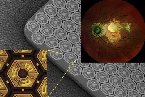
Fig. 9: PRIMA, a wireless subretinal implant made of photovoltaic pixels (Source: Pixium Vision). 2025 to today
Intracortical arrays that can both stimulate and record are being used to steer stimulation by the brain's response. Eduardo Fernández's group in Elche recorded neural activity around stimulating electrodes in two blind volunteers and showed that their visual experience could be predicted from the recorded activity [47]. Working with the groups of Michael Beyeler and Shih-Chii Liu (opens in a new tab), the same team then used deep learning to control stimulation-evoked population activity in blind human visual cortex. Their approach reached target responses at lower currents and elicited more consistent percepts than conventional methods [48].
Where the Field Stands
No visual prosthesis yet restores natural sight. But the field is broader today than at any point in its history. Cortical, retinal, optic nerve, and optogenetic approaches are being pursued in parallel, each suited to different causes of blindness. The open questions are less about whether artificial vision is possible than about how to make it reliable [49], useful in daily life [50], and sustainable for the people who receive it, including long-term support for the devices they carry.
Article information
How verification worksLast updated October 31, 2026 at 12:00 AM.
The following people contributed to this page:
- Michael BeyelerAssociate Professor, UC Santa Barbara